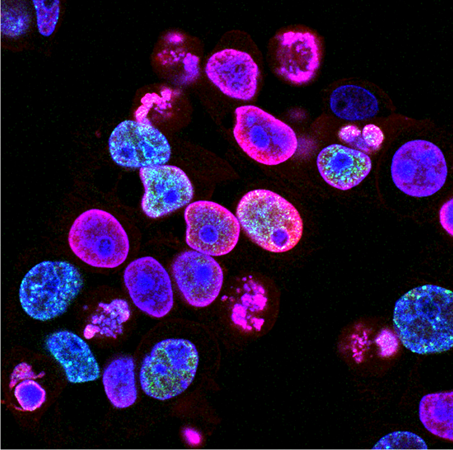
Custom Tagged Knock-In Immortalized & iPS Cells
Custom HiBiT-tagged knock-in cell lines for scalable, real-time endogenous protein quantification, visualization, and degradation studies.
Scalable, real-time endogenous protein analysis made simple
- Live-cell compatible: Track dynamic, real-time cellular processes without perturbing native protein function
- High sensitivity & specificity: Accurate quantification of low-abundance targets with minimal background
- Versatile applications: Adaptable for diverse experimental designs and research needs
- Streamlined workflows: Optimized to reduce assay complexity and variability for reproducible high-content or high-throughput studies
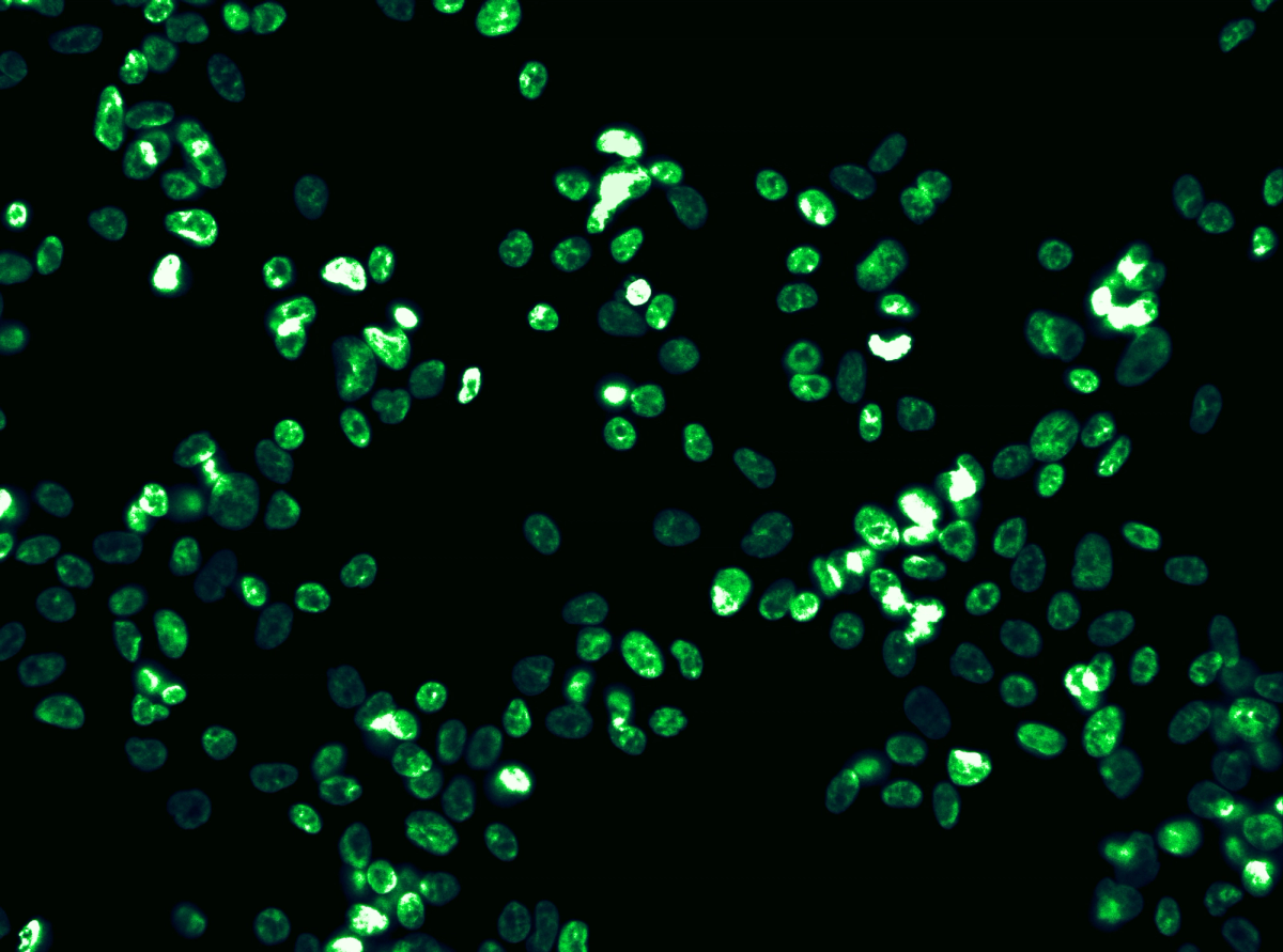
Cells available both clonally or as highly efficient pools with >70% average knock-in efficiency
HaloTagExplore More with HaloTag® Knock-ins
Multi-functional Tagging for Dynamic Biology
HaloTag® is a self-labeling protein tag that covalently binds to synthetic ligands, enabling precise control over how — and when — you visualize or manipulate a protein. By inserting HaloTag at the endogenous gene locus with CRISPR, EditCo gives you native expression and real-time control of your protein’s behavior, interactions, and location — without disrupting function. From high-resolution microscopy to pull-down assays and degradation studies, HaloTag is a powerful tool for studying protein fate across diverse cellular environments.
Why Choose HaloTag?
- Live-cell compatible — tag proteins with fluorescent dyes for real-time imaging
- Covalent and customizable — bind to ligands for imaging, purification, or degradation
- Highly specific — minimal background and no cross-reactivity with native proteins
- Dual-function — combine detection and capture in the same cell line
- Protein fate control — pair with HaloPROTACs or dTAGs for conditional protein degradation
Visualize Protein Localization: Track where and when your protein moves within the cell using fluorescent ligands in live imaging.
Study Protein-Protein Interactions: Use HaloTag pull-downs to identify complex members or binding partners under native conditions.
Enable Protein Purification: Capture your endogenously expressed protein of interest from native lysates using resin-based affinity ligands.
Quantify Trafficking and Turnover: Perform pulse-chase experiments with time-controlled ligand binding to monitor internalization and degradation.
When to Use HaloTag (vs. HiBiT or NanoLuc)
| Use Case | Optimal Tag |
|---|---|
| Live-cell imaging | HaloTag |
| Antibody-free protein quantification | HiBiT / NanoLuc |
| Pulse-chase protein turnover | HaloTag |
| Receptor internalization | HiBiT / NanoLuc |
| Pull-down or affinity purification | HaloTag |
| Conditional protein degradation | HaloTag |
| High-throughput reporter assay | HiBiT / NanoLuc |
Multiplex with Immunocytochemistry
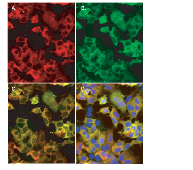
HaloTag® fusion proteins can be visualized alongside other cellular targets by combining fluorescent HaloTag ligands with immunocytochemistry. After labeling live cells with a HaloTag-compatible fluorophore, cells can be fixed and stained using an Anti-HaloTag polyclonal antibody paired with a spectrally distinct secondary antibody. This dual labeling approach helps validate in vivo ligand binding and ensures signal specificity. Because the antibody recognizes only the HaloTag protein, background staining is minimized, enabling clean, multiplexed imaging in fixed samples.
Study Membrane Protein Localization and Trafficking
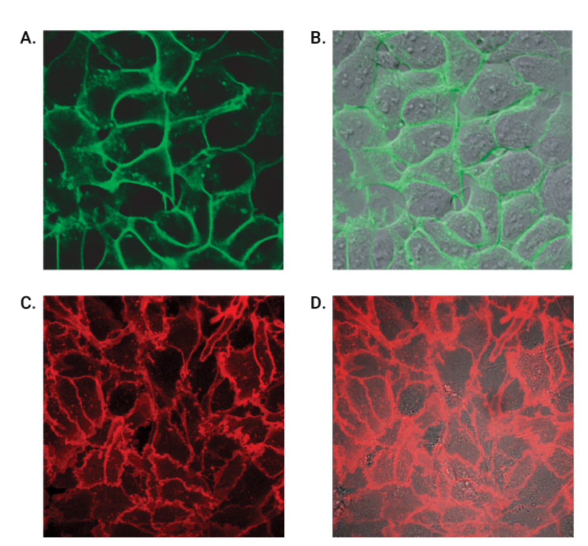
Track the localization and internalization of membrane-bound proteins by fusing them to HaloTag® and labeling with a fluorescent ligand that remains outside the cell. Since these ligands are cell-impermeant, they selectively bind only to surface-expressed proteins. The covalent interaction between the HaloTag protein and its ligand ensures the signal remains stable, even after fixation or under harsh experimental conditions. This allows you to confidently distinguish between surface-retained and internalized receptors, enabling detailed analysis of protein trafficking and membrane dynamics.
Achieve Superior Sensitivity with NanoLuc® Knock-ins
Ultra-Bright, ATP-Independent Luciferase for Sensitive, Scalable Detection
NanoLuc® luciferase is a next-generation, small (19.1 kDa) luciferase engineered for unparalleled luminescent performance. With >100-fold brightness compared to firefly or Renilla luciferase, NanoLuc enables high-sensitivity detection of low-abundance proteins and pathway activation — even in challenging biological systems.
When inserted at the endogenous locus via EditCo’s CRISPR knock-in workflow, NanoLuc becomes a native reporter, giving you a clean, quantitative signal without overexpression artifacts or the need for complex detection reagents.
Why Choose NanoLuc?
- Exceptional Brightness — delivers ultra-sensitive detection, even in low-expression systems
- Stable, Glow-Type Kinetics — ideal for endpoint or kinetic assays
- ATP-Independent Chemistry — less affected by cell health or energy status
- Small Size, Low Interference — preserves native protein function and dynamics
- Flexible Formats — supports secreted, intracellular, or split-luciferase (NanoBiT) designs
Integrating our core CRISPR expertise, high-quality reagents, and automated processes, we deliver the best edited cell-based models at any scale.